 |
Case Series
Transversus abdominis release for difficult ventral hernias – An open challenge for open abdomen
1 Post Graduate Resident, Institute of Surgical Gastroenterology & Liver Transplant, Govt. Stanley, Medical College, Tamil Nadu, Chennai, India
2 Senior Assistant Professor, Institute of Surgical Gastroenterology & Liver Transplant, Govt. Stanley Medical College, Tamil Nadu, Chennai, India
3 Assistant Professor, Institute of Surgical Gastroenterology & Liver Transplant, Tamil Nadu, Chennai, India
4 Professor, Institute of Surgical Gastroenterology & Liver Transplant, Govt. Stanley Medical College, Tamil Nadu, Chennai, India
5 Professor & Head of Department, Institute of Surgical Gastroenterology & Liver Transplant, Govt. Stanley Medical College, Tamil Nadu, Chennai, India
Address correspondence to:
Sugi R. V. Subramaniam
601, Old Jail Road,Institute of Surgical Gastroenterology & Liver Transplant, Govt. Stanley Medical College, Chennai, Royapuram, Tamil Nadu,
600001 India
Message to Corresponding Author
Article ID: 100030S05SS2018
Access full text article on other devices

Access PDF of article on other devices


How to cite this article
Sugi RV, Senthil Kumar P, Prabhakaran R, Kamalakannan R, Saravanan J, Thiruvarul M, Sugumar C, Jeswanth S, Ravichandran P. Transversus abdominis release for difficult ventral hernias – An open challenge for open abdomen. Edorium J Surg 2018;5:100030S05SS2018.ABSTRACT
Massive incisional hernias and open abdomen are serious challenges which the patient and surgeon face. Even more complicated are non-midline hernias, parastomal hernias, hernias near bony landmarks, and recurrent ventral hernias especially after anterior component separation. The available reconstructive techniques may not be sufficient to tackle this large defect to re-establish the lost abdominal domain and to create a long lasting successful tension free repair. The traditional approaches are haunted by serious complications of wound infection, skin necrosis and high incidence of recurrences. Posterior component separation with transversus abdominis release is a recent innovation of Classic Stoppa’s repair that offers a durable solution to a variety of complex ventral hernias. We report our initial experience with two cases of incisional hernias post multiple laparotomies and large midline rectus defects. Both had an uneventful post-operative course and were discharged without any wound related complications. They are on regular follow up outpatient visits and no recurrences were reported till date. This novel approach of posterior component separation with transversus abdominis release proves to be safe, effective and durable technique for difficult abdominal closure which in future can reduce the incidence of open abdomen to bare minimum.
Keywords: Posterior component separation, Sublay mesh, Transversus abdominis release
INTRODUCTION
Ventral hernias especially large incisional hernias are a frequent and challenging problem a surgeon encounters. They complicate about 11–23% of all abdominal surgeries [1]. The failure rates for various form of hernia repair ranges from 25–54% for primary suture repair, and up to 32% for open mesh repair [2]. Significant comorbidities, loss of domain and difficult anatomic considerations make management of massive recurrent hernias difficult. In patients undergoing multiple laparotomies for various indications it is not always easy to achieve abdominal closure, and open abdomen becomes inevitable. Open abdomen is associated with multiple morbidities like fistulisation, bleeding, infection etc [3]. Though there are multiple methods of achieving abdominal closure in difficult cases, the ideal method of hernia repair has not yet been found. Non-midline hernias, parastomal hernias, and recurrent ventral hernias after traditional anterior component separation present a greater challenge to repair. Typical reconstructive techniques in these scenarios would struggle to reestablish abdominal domain to create a lasting repair. We discuss two cases of massive incisional hernias in the background of multiple laparotomies in the past and how this novel approach of Transversus abdominis release helped in attaining tension free durable closure.
CASE SERIES
Case 1
A 54-year-old male, agriculture labourer presented with history of multiple laparotomies, operated elsewhere presented with complaints of protuberant abdomen. He did not have any complaints of altered bowel and bladder habits. He did not have any other specific complaints. On evaluation he gave history of multiple laparotomies within the past one year. A year ago he underwent emergency sigmoidectomy and covering ileostomy for sigmoid volvulus with gangrene. Two weeks later he underwent relaparotomy, adhesiolysis and primary closure of abdominal wall for acute abdomen. Six weeks later elective Ileostomy reversal was done. Unfortunately he developed anastomotic leak for which relaparotomy and resection with fresh anastomosis was done two days later. This time abdominal closure could not be achieved and he was managed as open abdomen with daily dressings and vacuum assisted negative pressure wound therapy. Once the wound granulated well he underwent split skin graft covering of the abdominal wound. He continued to have protruberant abdomen with visible bowel peristalsis. He was reassured and advised to wear abdomen corset. On examination his general condition was good. His abdomen was scaphoid on lying supine with split skin graft covering the elliptical midline defect which measured about 10 cm in the middle.Visible bowel peristalsis were seen. Other system examinations were normal. Routine laboratory investigations were normal and he did not have any comorbid illness. Abdominal imaging confirmed the inspector findings. He was planned for surgical exploration. On exploration, a defect in midline for about 8 cm in width was noted. Rectus was retracted laterally and could not be brought easily to the midline.Adhesiolysis was done. It was decided to go ahead with posterior component separation with Transverse abdominis release [TAR] since the defect was very wide. Posterior rectus sheath seperated from the anterior rectus at about 1 cm from the midline where the previous linea alba was present. Retrorectus dissection done till the level of linea semilunaris. Care was taken to preserve the neurovascular bundles encountered. Incision made on internal oblique fascia and the transverse abdominis muscle hooked and divided using an electrocautery. The transverse abdominis muscle fibres were released along its entire insertion line at the level of semilunaris extending from xiphoid process above. Inferiorly, it was separated till the level of arcuate semilunaris below which the muscles were deficient and only peritoneum was present. Laterally the release process was extended till bilateral psoas muscles were visualized. Superiorly it was extended till the central tendon of diaphragm. The posterior rectus sheath was approximated in midline using non absorbable sutures after placement of intra peritoneal drains. Polypropylene mesh of size approximately 30x15 cms were placed over the posterior rectus sheath covering in a sublay fashion and secured. Suction drain tubes were placed over the mesh covering and anterior rectus sheath approximated in midline without tension. Skin closed in midline Figure 1 (A–J). Daily vitals and drain output was monitored. After considerable decrease in drain output it was removed on fourth post-operative day. Abdominal sutures were removed at the end of second post-operative week. He was subsequently discharged a week later and was put on abdominal corset. Patient attended his routine outpatient visit after 2 weeks with no complaints.
Case 2
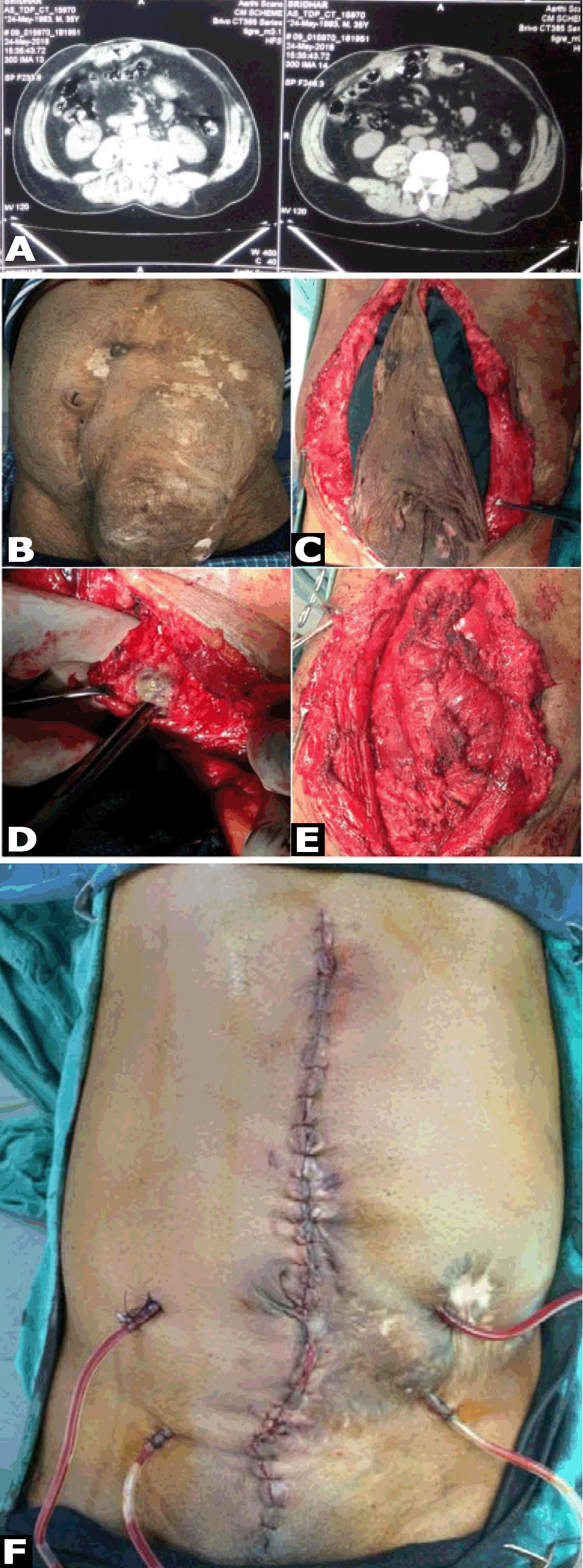
A 48-year-old male, a cook presented with similar history of multiple laparotomies presented with complaints of a bulge in the abdomen along previous scar site. His only complaint was the need to wear a corset round the clock and had a bulge on sitting up. Five years ago he underwent emergency Hartmann’s procedure for sigmoid transaction following blunt abdominal trauma. A month later he underwent colostomy reversal. He developed incisional hernia two months later for which meshplasty was done. He developed mesh infection following which he underwent re exploration and removal of infected mesh. Then he was managed as open abdomen for two months which after granulation was managed with split skin graft covering of the abdominal wound. He continued to have protruberant abdomen with a bulge which was more prominent on sitting up. He was forced to wear corset round the clock. On examination, his general condition was good. There was a bulge of size approximately 25x10 cms projecting from his anterior abdominal wall at the site of previous scar. A palpable midline rectus defect of 8 cm was noted. Visible bowel peristalsis were seen. Other system examinations were normal. Routine laboratory investigations were normal and he did not have any comorbid illness. Computed tomography of the abdomen confirmed thinning of the rectus sheath with focal outpouching of rectus in the infra umbilical region and herniation of small bowel loops (Figure 2A). He was planned for posterior component separation with transversus abdominis release. The posterior rectus was so thinned out with multiple inadvertent openings which were closed with absorbable sutures. Mesh placement was deferred owing to multiple openings in posterior rectus sheath and previous history of mesh infection requiring removal. Anterior rectus sheath was closed as a separate layer with non-absorbable sutures Figure 2(B–E). Suction drains were placed over the anterior rectus covering and skin closed over this. (Figure 2F) He was discharged after an uneventful post-operative course after two weeks.
DISCUSSION
The Classic Rives- Stoppa hernia repair, described way back in 1970s, uses the potential space between the posterior rectus fascia and the rectus muscle. This technique has been time tested and proven to be an effective approach for open ventral hernia repairs [4]. This allows placement of prosthetic mesh that extends approximately 6 to 8 cm on either side of the midline [5]. However, there are certain circumstances where the defect is greater and Stoppa’s repair alone is not sufficient to achieve a tension free midline approximation. Novitsky in 2012 introduced this new reconstructive technique of transversus abdominis release [TAR] and showed durable results with this technique [6]. This technique is considered as a modification of Stoppa’s GPRVS Giant Prosthetic Reinforcement of Visceral Sac] [7]. The extended retro rectus plane bounded by the psoas muscle laterally, central tendon of the diaphragm under the costal margin superolaterally, the inguinal ligament inferolaterally, and space of Retzius inferiorly [8]. This plane is utilized for mesh reinforcement and also to achieve a tension free midline rectus closure.
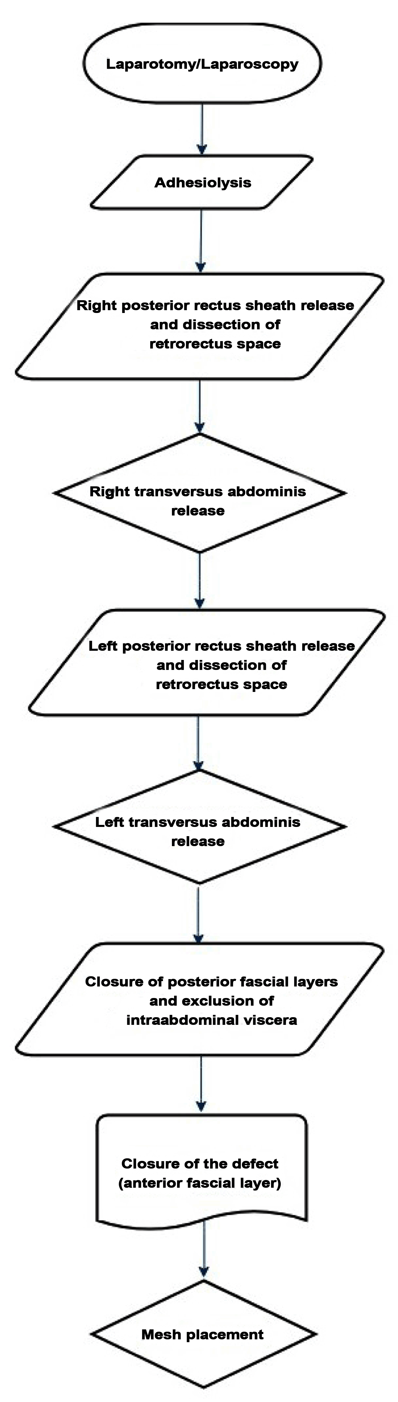
The main steps of the procedure are the same whether done by open or laparoscopic method (Flowchart 1). These include adhesiolysis, posterior rectus sheath release and dissection of retrorectus space on one side, transverse abdominis release following which contralateral posterior rectus sheath release and dissection of retrorectus space is done. Opposite side transversus abdominis release is carried out and closure of posterior fascial layers and exclusion of intraabdominal viscera is key step to avoid bowel injury. Closure of the anterior fascial layer. Mesh placement can be inlay, sublay or onlay although sublay is the most commonly employed technique.
Transversus abdominis release has several advantages which includes significant medial mobilization of the posterior rectus sheath that allows for extensive lateral dissection in a potentially unlimited space between the transversus muscle and the underlying transversalis fascia. It avoids disruption of the nerves and blood supply to the rectus abdominis and anterolateral abdominal wall skin thereby avoiding skin necrosis. Even though the exact clinical significance of preserving these neurovascular bundles remains debated, damage to the bundles has been suggested to predispose to abdominal wall bulging and laxity [9]. The combined contraction of the transverses abdominis and the posterior fibers of the internal oblique produce a tension around the abdominal wall similar to the one produced by a tight abdominal corset throughout the thoracolumbar fascia [10]. Key to successful abdominal wall repair is the strength of the rectus muscle.The rectus abdominis is innervated by the thoracoabdominal nerves T7 to T12, with additional supply from the lateral cutaneous nerve branch of T12, as well as the ilioinguinal and iliohypogastric (L1). The ilioinguinal and iliohypogastric nerves enter the space between the internal oblique and transversus abdominis via the lateral border of the transverses abdominis muscle. These nerves, along with the ventral primary rami of the lower six thoracoabdominal nerves (T7–T12) innervate the anterolateral abdominal wall skin and musculature [11]. The anterior abdominal wall receives its blood supply from the superior and inferior epigastric arteries, as well as the lower 6 intercostal arteries. These neurovascular bundles enter pass inferomedially between the internal oblique and transverses abdominis muscles before entering the lateral border of the rectus sheath [12].
Basically this technique potentially restores native biomechanics of the abdominal wall. On the long run TAR has proven to be versatile, reliable, durable and with low recurrence rate (3.7%) [13]. It is an easy-to-learn technique of hernia repair which once mastered can obviate the need for open abdomen and its complications. Laparoscopic abdominal wall reconstruction [AWR] with transversus abdominis release has proven to be an unique and feasible approach to complex abdominal wall defects with added benefits of laparoscopy such as reduced pain,early recovery, and lesser length of hospital stay [14]. More recently robotic assisted component separation techniques have been utilised for primary fascial defect closure [15].
CONCLUSION
This novel technique of abdominal wall reconstruction using a transversus abdominis muscle release is a modification of a posterior component separation and is an adjunct to the traditional retromuscular repair of Rives-Stoppa. TAR was associated with low perioperative morbidity, wound related complications and a low-recurrence rate. Posterior component separation via TAR appears to be a safe and effective method of ventral hernia repair, which should be a valuable addition to the armamentarium of surgeons performing complex abdominal wall reconstructions. TAR can also be done in patients with recurrent incisional hernias post anterior component separation, which can also be done via minimally invasive techniques like laparoscopic and robot assisted TAR.
REFERENCES
1.
Mudge M, Hughes LE. Incisional hernia: A 10 year prospective study of incidence and attitudes. Br J Surg 1985 Jan;72(1):70–1. [CrossRef]
[Pubmed]

2.
Flum DR, Horvath K, Koepsell T. Have outcomes of incisional hernia repair improved with time? A population-based analysis. Ann Surg 2003 Jan;237(1):129–35. [CrossRef]
[Pubmed]
3.
Kritayakirana K, M Maggio P, Brundage S, Purtill MA, Staudenmayer K, A Spain D. Outcomes and complications of open abdomen technique for managing non-trauma patients. J Emerg Trauma Shock 2010 Apr;3(2):118–22. [CrossRef]
[Pubmed]
4.
Novitsky YW, Porter JR, Rucho ZC, et al. Open preperitoneal retrofascial mesh repair for multiply recurrent ventral incisional hernias. J Am Coll Surg 2006 Sep;203 (3):283–9. [CrossRef]
[Pubmed]
5.
Rives J, Pire JC, Flament JB, Palot JP, Body C. Treatment of large eventrations. New therapeutic indications apropos of 322 cases. [Article in French]. Chirurgie 1985;111(3):215–25.
[Pubmed]
6.
Novitsky YW, Elliott HL, Orenstein SB, Rosen MJ. Transversus abdominis muscle release: A novel approach to posterior component separation during complex abdominal wall reconstruction. Am J Surg 2012 Nov;204(5):709–16. [CrossRef]
[Pubmed]
7.
Stoppa R, Petit J, Abourachid H, Henry X, Duclaye C, Monchaux G, Hillebrant JP. Original procedure of groin hernia repair: Interposition without fixation of Dacron tulle prosthesis by subperitoneal median approach. [Article in French]. Chirurgie 1973 Feb;99(2):119–23.
[Pubmed]
8.
Oprea V, Radu VG, Moga D. Transversus abdominis muscle release (TAR) for large incisional hernia repair. Chirurgia (Bucur) 2016 Nov-Dec;111(6):535–40. [CrossRef]
[Pubmed]
9.
Rosen MJ, Fatima J, Sarr MG. Repair of abdominal wall hernias with restoration of abdominal wall function. J Gastrointest Surg 2010 Jan;14(1):175–85. [CrossRef]
[Pubmed]
10.
Bogduk N, Twomey TL. Clinical Anatomy of the Lumbar Spine. 2ed. Philadelphia, PA: Churchill Livingstone; 1991.
11.
Moore K. Clinically Oriented Anatomy. 5ed. Philadelphia, PA: Lipincott William & Wilkins; 2006.
12.
Hammond DL, Ackerman L, Holdsworth R, Elzey B. Effects of spinal nerve ligation on immunohistochemically identified neurons in the L4 and L5 dorsal root ganglia of the rat. J Comp Neurol 2004 Aug 2;475(4):575–89. [CrossRef]
[Pubmed]
13.
Novitsky YW, Fayezizadeh M, Majumder A, Neupane R, Elliott HL, Orenstein SB. Outcomes of posterior component separation with transversus abdominis muscle release and synthetic mesh sublay reinforcement. Ann Surg 2016 Aug;264(2):226–32. [CrossRef]
[Pubmed]
14.
Belyansky I, Zahiri HR, Park A. Laparoscopic transversus abdominis release, a novel minimally invasive approach to complex abdominal wall reconstruction. Surg Innov 2016 Apr;23(2):134–41. [CrossRef]
[Pubmed]
15.
Schluender S, Conrad J, Divino CM, Gurland B. Robot-assisted laparoscopic repair of ventral hernia with intracorporeal suturing. Surg Endosc 2003 Sep;17(9):1391–5. [CrossRef]
[Pubmed]
SUPPORTING INFORMATION
Author Contributions
Sugi R. V. Subramaniam - Substantial contributions to conception and design, Acquisition of data, Analysis of data, Interpretation of data, Drafting the article, Revising it critically for important intellectual content, Final approval of the version to be published
Senthil Kumar Perumal - Substantial contributions to conception and design, Acquisition of data, Analysis of data, Interpretation of data, Drafting the article, Revising it critically for important intellectual content, Final approval of the version to be published
Prabhakaran Raju - Substantial contributions to conception and design, Acquisition of data, Analysis of data, Interpretation of data, Drafting the article, Revising it critically for important intellectual content, Final approval of the version to be published
Kamalakannan Rajendran - Substantial contributions to conception and design, Acquisition of data, Analysis of data, Interpretation of data, Drafting the article, Revising it critically for important intellectual content, Final approval of the version to be published
Saravanan Janakiraman - Substantial contributions to conception and design, Acquisition of data, Analysis of data, Interpretation of data, Drafting the article, Revising it critically for important intellectual content, Final approval of the version to be published
Thiruvarul Muthukumarasamy - Substantial contributions to conception and design, Acquisition of data, Analysis of data, Interpretation of data, Drafting the article, Revising it critically for important intellectual content, Final approval of the version to be published
Jeswanth Sathyanesan - Substantial contributions to conception and design, Acquisition of data, Analysis of data, Interpretation of data, Drafting the article, Revising it critically for important intellectual content, Final approval of the version to be published
Ravichandran Palaniappan - Substantial contributions to conception and design, Acquisition of data, Analysis of data, Interpretation of data, Drafting the article, Revising it critically for important intellectual content, Final approval of the version to be published
Guaranter of SubmissionThe corresponding author is the guarantor of submission.
Source of SupportNone
Consent StatementWritten informed consent was obtained from the patient for publication of this case series.
Data AvailabilityAll relevant data are within the paper and its Supporting Information files.
Conflict of InterestAuthors declare no conflict of interest.
Copyright© 2018 Sugi R. V. Subramaniam et al. This article is distributed under the terms of Creative Commons Attribution License which permits unrestricted use, distribution and reproduction in any medium provided the original author(s) and original publisher are properly credited. Please see the copyright policy on the journal website for more information.


