 |
Case Report
Neuroprosthetic rehabilitation using titanium cranial prosthesis
1 MDS, Prosthodontis and Implantologis Ex Senior Resident Government Dental College, Thiruvananthapuram Consultant Bhaktivedanta Hospital Mira Road, Mumbai
2 MDS, Assistant Professor, Department of Oral Pathology and Microbiology, D.J. College of Dental Sciences and Research , Uttar Pradesh, Modinagar, Ghaziabad, India
Address correspondence to:
Muneet Marwah
MDS, Prosthodontis and Implantologis Ex Senior Resident Government Dental College, Thiruvananthapuram Consultant Bhaktivedanta Hospital Mira Road,
Mumbai
Message to Corresponding Author
Article ID: 100034S05MM2018
Access full text article on other devices

Access PDF of article on other devices


How to cite this article
Marwah M, Sekhon HK. Neuroprosthetic rehabilitation using titanium cranial prosthesis. Edorium J Surg 2018;5:100034S05MM2018.ABSTRACT
Cranioplasty is a surgical procedure to repair the cranial defect or deformation. The goal of cranioplasty is to attain the accurate closure of the defect by using various biocompatible materials there by improving their neurologic status and restoring the esthetics of the patient in a best possible way. One of the earliest materials used for restoring the cranial defects were autologous bone grafts. With the advancement and to overcome the disadvantages of autologous bone grafts many synthetic materials have been introduced. This article illustrates a case report of neuroprosthetic rehabilitation of a patient who sustained skull fracture after a road traffic accident followed by decompressive craniectomy and cranioplasty using titanium cranial prosthesis improving neurologic status and esthetics of the patient.
Keywords: Cranioplasty, Cranial prosthesis, Neuroprosthetic rehabilitation
INTRODUCTION
Defects in the cranial region can result due to trauma,disease or congenital malformations. Repair of these defects becomes necessary to protect brain tissue underneath, to relieve the pain at defect site, and improve aesthetics. Cranioplasty is a surgical procedure to repair the defects in the cranium. It usually is done after a craniectomy or a craniotomy [1]. In all age groups, the main reason for doing cranioplasty are either tumor removal or decompressive craniectomies following traumatic injuries. Different types of materials have been used to repair the cranial defects. An ideal material should have features such as, radiolucency, resistance to infection, must not change dimensionally due to heat, easy to shape, economical, resistant to biomechanical processes, adapts to the margins of the cranial defects properly and achieves complete closure [1],[2]. Although no such ideal material is available, but throughout history materials used were autologous bone, xenografts, allografts, various synthetic materials like metals, methyl methacrylate, hydroxyapetite, ceramics, titanium mesh, polyether ether ketone (PEEK), and Carbon-fibrereinforced polymer (CFRP) [2],[3]. Following cranioplasty improvement in electroencephalographic abnormalities, neurological dysfunction, seizures and cerebral blood flow abnormalities have been reported.
CASE REPORT
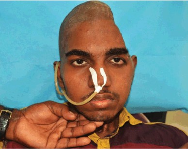
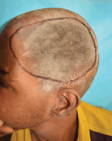
A 20-year-old male patient with history of road traffic accident suffered skull fracture for which he underwent decompressive craniectomy in the Neurosurgery department, Government Medical College, Thiruvananthpuram. Further, he was referred to prosthodontics department, Government Dental College Thiruvananthpuram for the prosthetic rehabilitation of the surgical defect. It involved frontal, parietal and temporal bone (Figure 1), (Figure 2).
Impression procedure
It requires patient’s scalp to be completely shaven before making the impression. Margins of the defect are then palpated to determine the periphery of the defect,they are traced with an indelible pencil and transferred in the impression. Modeling wax was adapted to the margins of the defect to act as a boundary for the impression material. A loose smooth mix of alginate hydrocolloid material was poured on the defect area avoiding air entrapment. Tuft of cotton were placed over the alginate material before it completely sets. This acted as a binder between alginate and a loose layer of Plaster of Paris that was poured over it. Plaster of Paris supported the impression material giving it strength and preventing its distortion during removal. Impression was poured with dental stone (Kalstone, Kalabhai, Mumbai, India) to obtain a positive replica of the patient’s skull. Markings to delineate the margins of the defect were not as precise as originally placed so the centre of the line was assumed to be the border of the defect. The design and the extent of the prosthesis was discussed with the neurosurgeon before proceeding for the final fabrication of the cranial prosthesis (Figure 3).
Wax pattern trial
With the help of positive replica of patient’s skull (stone cast), a wax pattern was made on the defect area to the exact dimensions and contour simulating the contra lateral normal side. It was tried on the patient and the outline of wax pattern was checked and modified from all sides frontal, saggital and occipital to restore the normal contour of the patient’s skull. The margins were beveled to ensure proper adaptation of wax prosthesis with the defect area (Figure 4), (Figure 5).
Titatnium plate fabrication
The wax pattern was invested, a mould formed and a pure titanium sheet (medical grade) was used to fabricate a titanium plate (Jayon Implant Pvt Ltd. Lab, Kanjikode, Palakkad, Kerala) (Figure 6). Holes (2 mm in diameter) were drilled throughout the plate that helped to reduce the chances of an epidural hematoma, allowed in-growth of fibrous connective tissue to assist in stabilization and permitted escape of underlying fluid and its absorption by the lymphatics of the scalp (Figure 7). The holes must be counter sunk on both inner and outer surface to prevent its sharp margins from cutting the sutures. They also help in securing the cranial prosthesis to the bony defect. The titanium plate was then autoclaved and made ready for the surgical procedure.
Surgical procedure for placement of titanium plate
Oro-tracheal intubation was done after keeping the patient in a supine patient. The incision was planned after considering need for wide exposure, tension free closure and a concealed position for the final scar. Also, it was marked over healthy tissue as required ideally far from the area of reconstruction. After starting the intravenous administration of antibiotics, cutaneous incision was opened in layers and dissection was done bluntly in subgaleal plane without disrupting the pericranium. Incision was then retracted with opposing skin hooks and dissection was carried further till the entire defect margins were delineated in a supraperiosteal plane. Using electrocautery marking were made on the intact bony margin corresponding to the holes on the periphery of the titanium plate. Holes were drilled into the diploe layer of the bone at the previously marked points through the outer table of the skull using no 40 bone bur under copious saline irrigation. Another hole was drilled into the diploe layer contacting each of the former holes at the right angle. A proline suture (1–0) was passed through all these holes giving tripod stability to the plate. After the initial stabilization plate was evaluated for contour, position and bulk and finally sutured firmly to the preferred area. The pericranium and galea are approximated and closure was done in layers using vicryl. Skin was sutured using staple sutures with a drain placed and left in place along with compressive turban dressing (Figure 8), (Figure 9), (Figure 10), (Figure11). Postoperatively, antibiotics were continued for five days. After one week the patient was recalled and examined and it was observed that there was a remarkable improvement in the contour of the skull.
DISCUSSION
Cranioplasty is most commonly performed after craniectomy or craniotomy in cases following severe head injuries. Other indications of cranioplasty being refractory intracranial hypertension, large vessel infarct, intraopeative brain swelling,encephalitis and aneurysmal subarachnoid hemorrhage. Cranioplasties conducted one to six months after craniectomy have the highest complication rates (7.9%) and those performed twelve to eighteen months after craniectomy have the lowest complication rates (4.5%) [4],[5],[6],[7]. The syndrome of trephined as described by Gardner is accepted as an indication to reconstruct the skull. Its features include dizziness, headache, irritability, intolerance, fatiguability, loss of motivation, depression and anxiety. There are evidences of improvement in symptoms after cranioplasty with the reversion of intracranial pressure relationships to normal. Improvements in neurologic dysfunction, seizures, electroencephalographic abnormalities have been reported [8],[9],[10]. Contraindications for performing cranioplasty include cerebral swelling, compound wound, infection, hydrocephalus, functional paranasal sinuses, and scarred ,thin or devitalized scalp.
Cranioplasty as a neurosurgical procedure dates back to 3000 BC [11]. A range of biomaterials including bone autografts, xenografts, allografts, celluloids, polymethylmethacrylate, metals such as aluminum, gold, silver, and titanium, polyethylene, silicon, and many others [11] have been used to develop an adequate topographical substitute for a cranial defect. Autogenous bone has been historically preferred over alloplastic materials to reconstruct the cranium, due to allegedly better mechanical, biologic, and immunologic properties [12],[13]. Its available in the form of patient’s own bone flap or by bone harvesting. Harvesting a bone flap requires additional surgeries and has associated morbidities because of which its use has been reduced over the years. While using the original bone flap the two main problems are not having the complete flap available due to initial trauma or if it is available the main problem is its storage. The most common method is to freeze the bone and store it in a bone bank. It keeps the bone matrix architecture intact but causes tissues to die resulting in partial resorption. Another method to store craniectomy flap is to store it in fatty tissues of the abdomen. This method also has certain disadvantages like it causes an additional surgical wound, chances of infection and results in abdominal scar. Autologous bone grafts have the highest rate of infection (25.9%) compared to polymethylmethacrylate (PMMA), alumina ceramics and titanium mesh [14]. In cases where we cannot use autogenous bone grafts alloplastic materials like PMMA and titanium are viable alternatives most commonly used today [12],[15]. Methyl methacrylate was first used in 1940 by Zander as a cranioplastic material [4]. It has many advantages like its chemically inert, light weight, nonmagnetic, non thermoconductive and comparable to bone in strength [16]. Its malleable before it sets so therefore gives good cosmetic results. But its main drawback is that during the setting of polymer it produces excessive heat due to its exothermic reaction that can damage the underlying brain tissue. It also shatters easily because of its brittle nature. Blum et al reported a complication rate of 23% within eight years of surgery after studying long term consequences of MMA usage for cranioplasty, complications mainly being infections [17].
Therefore, it was planned to use the next best available material titanium as a material of choice for the restoration of skull defect. Titanium is a noncorrosive metallic alloy, with low risk of infection that provides superb cosmetic results. It has high overall strength and malleability. Matsuno reported that of all cranioplasty material titanium mesh has lowest rate of graft infection (2.6%) [18]. It shows no toxicity, has no inflammatory reaction and allows bone growth into its porous spaces and through openings of its mesh like structure. Another advantage is the accurate postoperative imaging without any major artifacts exploring the CT or MRI. The main disadvantage is that it is difficult to mold intraoperatively and has high cost.
Recent advances
PEEK implants are commonly used today because with 3D printing technologies they can be designed specific to a patient’s craniotomy defect. Computer assisted 3D modelling can be used to design these synthetic implants. They are radiolucent, chemically inert, and can be sterilized with steam or gamma irradiation [19]. They have thickness, strength and elasticity comparable to cortical bone and can be incorporated accurately within the defect without use of miniplates. The disadvantages are it lacks osteointegrative properties and are expensive. There is lack of sufficient literature on the risk of infections with peek implants and needs further research.
Future of cranioplasty
With the help of recent technology there is an ongoing research on both biologic and non-biologic substitutes. Preforming of implants has advanced due to 3D-CT scanning, steriolithography [20], and computer assisted design [21]. Development in tissue engeneering using molecular biology techniques like mesenchymal stem cell or harvesting osteoblasts to seed onto the scaffold for the cranioplasty are taking place [22]. In future biodegradable implants may be used in which releasing bioactive molecules will transform perfectly fitted implant into living bone providing immediate cover of the cranial defect.
CONCLUSION
Due to a large number of cranial injuries occurring in this modern age the number of patients requiring cranioplasty has gone up to a large number. Till now there is no ideal material for cranioplasty, but materials that are strong, inexpensive, resistant to infection, radiolucent and able to reincorporate with a patient’s craniotomy defect will give greatest advantages for such patients. A case report of a patient has been illustrated who had a road traffic accident with multiple fractures including skull followed by decompressive craniectomy and a cranioplasty rehabilitating his neurological status and cosmesis using a titanium cranial prosthesis. Though the conventional method was used for titanium cranial prosthesis fabrication but satisfactory results both esthetically and functionally were obtained.
REFERENCES
1.
Shah AM, Jung H, Skirboll S. Materials used in cranioplasty: A history and analysis. Neurosurg Focus 2014;36(4):1–7. [CrossRef]
[Pubmed]

2.
Aydin S, Kucukyuruk B, Abuzayed B, Aydin S, Sanus GZ. Cranioplasty: Review of materials and techniques. J Neurosci Rural Pract 2011;2(2):162–7. [CrossRef]
[Pubmed]
3.
Höhne J, Brawanski A, Gassner HG, Schebesch KM. Feasibility of the custom-made titanium cranioplasty CRANIOTOP(®). Surg Neurol Int 2013;4:88. [CrossRef]
[Pubmed]
4.
Beumer J 3rd, Curtis TA, Marunick MT. Maxillofacial rehabilitation: Prosthodontic and surgical considerations. St. Louis: Ishiyaku EuroAmerica; 1996. p. 455–5.
5.
Jordan RD, White JT, Schupper N. Technique for cranioplasty prosthesis fabrication. J Prosthet Dent August 1978;40(2)230–. [CrossRef]
[Pubmed]
6.
Jaskolka MS, Olavarria G. Reconstruction of skull defects. Atlas Oral Maxillofac Surg Clin North Am 2010;18(2):139–49. [CrossRef]
[Pubmed]
7.
Rish BL, Dillon JD, Meirowsky AM, et al. Cranioplasty: A review of 1030 cases of penetrating head injury. Neurosurgery 1979;4(5):381–85.
[Pubmed]
8.
Grantham EG, Landis HP. Cranioplasty and the post-traumatic syndrome. J Neurosurg 1948;5(1):19–22. [CrossRef]
[Pubmed]
9.
Rifkinson-Mann S. Cranial surgery in ancient Peru. Neurosurgery 1988;23(4):411-6.
[Pubmed]
10.
Kent JN, Zide MF. Wound healing: Bone and biomaterials. Otolaryngol Clin North Am 1984:17(2):273–319.
[Pubmed]
11.
Sanan A, Haines SJ. Repairing holes in the head: A history of cranioplasty. Neurosurgery 1997;40(3):588–603.
[Pubmed]
12.
Marchac D, Greensmith A. Long term experience with methylmethacrylate cranioplasty in craniofacial surgery. J Plast Reconstr Aesthet Surg 2008;61(7):744–53. [CrossRef]
[Pubmed]
13.
Sahoo N, Roy ID, Desai AP, Gupta V. Comparative evaluation of autogenous calvarial bone graft and alloplastic materials for secondary reconstruction of cranial defects. J Craniofac Surg 2010;21(1):79–82. [CrossRef]
[Pubmed]
14.
Matsuno A, Tanaka H, Iwamuro H, et al. Analyses of the factors influencing bone graft infection after delayed cranioplasty. Acta Neurochir(Wien) 2006;148(5):535–40. [CrossRef]
[Pubmed]
15.
Moreira Gonzalez A, Jackson IT, Miyawaki T, Barakat K, DiNick V. Clinical outcome in cranioplasty: Critical review in long term follow up. J Craniofac Surg 2003;14(2):144–53.
[Pubmed]
16.
Winn HR, Youmans JR. Youmans Neurological Surgery. 3ed. Philadelphia: Saunders; 1990.
17.
Blum KS, Schneider SJ, Rosenthal AD. Methyl methacrylate cranioplasty in children: Long-term results. Pediatr Neurosurg 1997;26(1):33–5. [CrossRef]
[Pubmed]
18.
Matsuno A, Tanaka H, Iwamuro H, et al. Analyses of the factors influencing bone graft infection after delayed cranioplasty. Acta Neurochir (Wien) 2006;148(5):535–40. [CrossRef]
[Pubmed]
19.
Lethaus B, Safi Y, ter Laak-Poort M, et al. Cranioplasty with customized titanium and PEEK implants in a mechanical stress model. J Neurotrauma 2002;29(6):1077–83. [CrossRef]
[Pubmed]
20.
Eppley B, Kilgo M, Coleman J. Cranial reconstruction with computer-generated hard-tisue replacement patient-matched implants: Indications, surgical technique and long-term follow-up. Plast Reconstr Surg 2002;109(3):864–71.
[Pubmed]
21.
Gelaude F, Vander Sloten J, Lauwers B. Automated design and production of cranioplasty plates: outer surface methodology, accuracies and a direct comparison to manual techniques. Computer-Aided Design and Applications 2006;3(1–4):193–202. [CrossRef]
22.
Chim H, Schantz J. New frontiers in calvarial reconstruction: Integrating computer-assisted design and tissue engineering in cranioplasty. Plast Reconstr Surg 2005;116(6):1726–41. [CrossRef]
[Pubmed]
SUPPORTING INFORMATION
Author Contributions
Muneet Marwah - Substantial contributions to conception and design, Acquisition of data, Analysis of data, Interpretation of data, Drafting the article, Revising it critically for important intellectual content, Final approval of the version to be published
Harjeet Kaur Sekhon - Substantial contributions to conception and design, Acquisition of data, Drafting the article, Revising it critically for important intellectual content, Final approval of the version to be published
Guaranter of SubmissionThe corresponding author is the guarantor of submission.
Source of SupportNone
Consent StatementWritten informed consent was obtained from the patient for publication of this case report.
Data AvailabilityAll relevant data are within the paper and its Supporting Information files.
Conflict of InterestAuthors declare no conflict of interest.
Copyright© 2018 Muneet Marwah et al. This article is distributed under the terms of Creative Commons Attribution License which permits unrestricted use, distribution and reproduction in any medium provided the original author(s) and original publisher are properly credited. Please see the copyright policy on the journal website for more information.
Comment on Article

