 |
Case Report
Spigelian hernia successfully managed with a laparoscopic approach, the first ever case reported in Ecuador
1 Attending Surgeon, Department of General Surgery, Hospital IESS Quito Sur, Quito, Ecuador
2 Chief of General Surgery, Department of General Surgery, Hospital IESS Quito Sur, Quito, Ecuador
3 Surgery Resident, Department of General Surgery, Hospital IESS Quito Sur, Quito, Ecuador
Address correspondence to:
Gabriel A Molina
MD, Attending Surgeon, Department of General Surgery, Hospital IESS Quito Sur, Quito,
Ecuador
Message to Corresponding Author
Article ID: 100043S05GF2020
Access full text article on other devices

Access PDF of article on other devices


How to cite this article
Fuentes G, Molina GA, Jiménez GE, Jiménez AS, Cedeño AB, Robles MA, Mosquera JM, Carrera PA, Hernández O. Spigelian hernia successfully managed with a laparoscopic approach, the first ever case reported in Ecuador. Edorium J Surg 2020;7:100043S05GF2020.ABSTRACT
Spigelian hernia is an abdominal wall defect through the Spigelian fascia. These hernias are relatively rare hernias that account for only 2% of all the abdominal wall hernias. Diagnosis is difficult as these hernias have nonspecific symptoms and a palpable mass is usually not identified. A high clinical awareness along with complementary exams are usually needed to confirm the diagnosis. Surgery is required as these hernias have a high risk of strangulation and incarceration. The advent of laparoscopic surgery has improved surgical outcomes. In developing countries the availability of this equipment is limited; however, adequate qualification and training of surgeons in these techniques are vital for achieving satisfactory results. We report the case of a 58-year-old patient with a Spigelian hernia. It was successfully managed through laparoscopy and it is the first case ever reported in Ecuador.
Keywords: Abdominal hernia, Hernia, Laparoscopy, Spigelian hernia
INTRODUCTION
Spigelian hernia is a protrusion of preperitoneal fat, or viscus through a congenital defect or weakness in the Spigelian fascia. These rare hernias are usually located along the semilunar line close to the level of the arcuate line [1],[2]. Diagnosis requires a high index of suspicion, given the lack of symptoms and signs [1],[3]. Clinical history, physical examination, and preoperative imaging are vital to achieve an accurate diagnosis. Surgery is necessary to repair the defect, but better and more reliable results are guaranteed with the help of laparoscopy [1],[4],[5]. We report the case of a 58-year-old patient with a Spigelian hernia. It was successfully managed through laparoscopy and it is the first case ever reported in Ecuador.
CASE REPORT
We report the case of a 58-year-old female with past medical history of bilateral inguinal hernia and bilateral salpingectomy. She presented to the emergency room with acute abdominal pain in her right abdomen. Her vital signs were normal; heart rate was 82 beats per minute, blood pressure was 148/88 mmHg, and her body mass index (BMI) was 22. She had a 1-year history of a growing mass in her lower right abdomen, which was initially small and gradually progressed to its current size. The mass increased in size when coughing but partially decreased when lying down. At first, the mass did not cause pain or discomfort, so she did not pay much attention to it. However, one month before her admission, she remembered that she suffered brief intermittent episodes (2–3 minutes approximately) of acute abdominal pain during exercise that resolved spontaneously.
On clinical examination a painful 4 × 4 cm mass was detected in her right abdomen, the mass was located in the lateral border of the rectus abdominis muscle and the semilunar line, 2 cm from the midline laterally and 3 cm cranially from the right anterior superior iliac spine. The skin around the mass was swollen and had multiple erythematous patches. We were unable to fully manipulate the mass as she had severe pain on palpation. Her abdomen was soft, but she had tenderness around the mass. The patient denied changes in bowel functions, discomfort on exercise, or vaginal discharge.
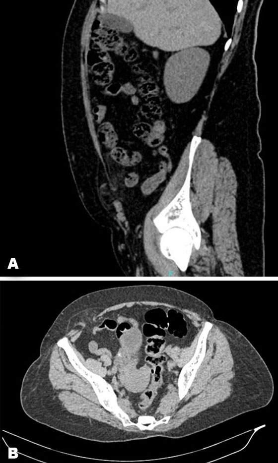
Complementary exams were requested, a complete blood count was normal and an abdominal echography was inconclusive. Due to this, an abdominal computed tomography (CT) revealed a 4 × 4 cm abdominal wall defect in the right semilunar line. The hernia had a segment of trapped omentum along with slight inflammatory changes, the loops of the small bowel appeared normal without signs of obstruction, an incarcerated Spigelian hernia was diagnosed (Figure 1A and Figure 1B).
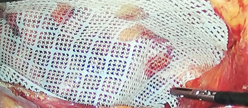
A laparoscopic approach was decided, three laparoscopic ports were used for the repair, an 11 mm umbilical port and two 5 mm ports. One at the left midclavicular line in the left upper quadrant and another at the right midclavicular line in the right upper quadrant. Multiple adhesions were seen between the omentum and the abdominal wall, which were removed using an ultrasound device (Harmonic, Ethicon, Johnson & Jhonson, Cincinnati, USA). The section of the omentum that was incarcerated was resected and the abdominal wall defect was visualized. The defect was repaired using a nonabsorbable suture and secondarily reinforced with a 7 × 5 cm polytetrafluoroethylene mesh (Proceed, Ethicon, Johnson & Jhonson, Somerville, NJ, USA) and secured using a suture passer, after this, the mesh was in place with an excellent overlap of the defect (Figure 2). The rest of the procedure was completed without complications.
Her postoperative course was uneventful, liquid diet was initiated on the first postoperative day and was followed by a full diet. She was discharged on her third postoperative day without complications. She was able to return to her exercise routine three months after surgery without any complications. One year after surgery she is doing well.
DISCUSSION
Spigelian hernia is named after Adriaan van den Spieghel who first described the semilunar line in 1645, but it was Josef Klingosh who reported this abdominal wall defect in 1764 [1],[2]. These hernias appear in an area limited medially by the lateral edge of the rectus muscle and laterally by the semilunar line [1],[2]. Most of the Spigelian hernias appear in the area below the umbilicus and above the interspinal plane [3]. This area is thought to be an area of weakness since there is a musculoaponeurotic transition lateral to the rectus muscle and the aponeuroses of the internal oblique and the transversus. Here, the aponeuroses change in direction and run parallel rather than at the angle found in the planes above the umbilicus [1],[3],[4]. Spigelian hernia is a rare event and accounts only for 0.12–2% of all abdominal wall hernias. Since the defect is covered by the aponeurosis of the external oblique, it can be difficult to palpate [1],[5]. Symptoms are usually vague and nonspecific, most patients present with abdominal pain, and few with a palpable mass in the lower abdomen, that is why most cases are diagnosed in an emergency setting [1],[6]. As it happened in our patient. Diagnosis is difficult and additional tests like ultrasonography or CT are usually needed [1],[3],[7]. Predisposing factors include obesity, collagen disorders, and chronic pulmonary obstructive disease and prior abdominal surgeries [1],[4]. In our case, the patient had a history of inguinal hernia and salpingectomy.
As the defect is usually narrow, it is predisposed to strangulation, this situation can occur in up to 21% of the cases. Spigelian hernias typically contain omentum, but small bowel, colon, appendix, bladder, and Meckel’s diverticulum have been reported. Therefore, surgery is recommended [1],[3]. In our patient, the omentum was found defective. The open approach has been the traditional approach. Treating the hernia and placing a mesh in a sublay or onlay position was the only treatment until 1992 when Carter and Mizes first described a laparoscopic approach with less postoperative pain, morbidity, and the ability to visualize the contralateral side [1],[3],[7]. Since then, most cases are now being managed taking advantage of minimal access surgery. Laparoscopic intraperitoneal onlay mesh (IPOM), laparoscopic total extraperitoneal technique (TEP), and laparoscopic transabdominal preperitoneal (TAPP) techniques are available. Nonetheless, due to the adhesion risk many surgeons opt for TAPP or TEP repair [1],[4].
Spigelian hernias are rare abdominal wall hernias that have a broad spectrum of clinical manifestations and wide differential diagnoses. High clinical awareness is necessary for an accurate diagnosis as incarceration and strangulation can lead to severe complications. Laparoscopic repair is an effective method of repair with excellent outcomes.
CONCLUSION
Spigelian hernias are rare events and their presentation is usually nonspecific. This makes diagnosis and prompt treatment troublesome. A high index of suspicion is needed as these hernias can quickly worsen and lead to dangerous complications. Also, in a unique way this case also proves that laparoscopic and surgical techniques along with optimal training should be a critical part for physicians, even in limited resource countries like our own changes in medical education are necessary to achieve better results.
REFERENCES
1.
Barnes TG, McWhinnie DL. Laparoscopic spigelian hernia repair: A systematic review. Surg Laparosc Endosc Percutan Tech 2016;26(4):265–70. [CrossRef]
[Pubmed]

2.
Kelly ME, Courtney D, McDermott FD, et al. Laparoscopic spigelian hernia repair: A series of 40 patients. Surg Laparosc Endosc Percutan Tech 2015;25(3):e86–9. [CrossRef]
[Pubmed]
3.
Moreno-Egea A, Carrasco L, Girela E, MartÃn JG, Aguayo JL, Canteras M. Open vs laparoscopic repair of spigelian hernia : A prospective randomized trial. Arch Surg 2002;137(11):1266–8. [CrossRef]
[Pubmed]
4.
Palanivelu C, Vijaykumar M, Jani KV, Rajan PS, Maheshkumaar GS, Rajapandian S. Laparoscopic transabdominal preperitoneal repair of spigelian hernia. JSLS  2006;10(2):193–8.
[Pubmed]
5.
Mederos R, Lamas JR, Alvarado J, Matos M, Padron I, Ramos A. Laparoscopic diagnosis and repair of Spigelian hernia: A case report and literature review. Int J Surg Case Rep 2017;31:184–7. [CrossRef]
[Pubmed]
6.
Filip S, Dragomirescu C, Copăescu C. Laparoscopic treatment of Spiegel hernia by total extraperitoneal (TEP) approach. Chirurgia (Bucur) 2014;109(3):325–9.
[Pubmed]
7.
Mittal T, Kumar V, Khullar R, Sharma A, Soni V, Baijal M, Chowbey PK. Diagnosis and management of Spigelian hernia: A review of literature and our experience. J Minim Access Surg 2008;4(4):95–8. [CrossRef]
[Pubmed]
SUPPORTING INFORMATION
Author Contributions
Germanico Fuentes - Substantial contributions to conception and design, Interpretation of data, Revising it critically for important intellectual content, Final approval of the version to be published
Gabriel A Molina - Substantial contributions to conception and design, Acquisition of data, Analysis of data, Interpretation of data, Drafting the article, Final approval of the version to be published
Galo Enrique Jimén - Substantial contributions to conception and design, Interpretation of data, Drafting the article, Final approval of the version to be published
Andres Sebastian Jiménez - Substantial contributions to conception and design, Interpretation of data, Revising it critically for important intellectual content, Final approval of the version to be published
Ana Belen Cedeño - Substantial contributions to conception and design, Interpretation of data, Drafting the article, Final approval of the version to be published
Marcela Alexandra Robles - Substantial contributions to conception and design, Interpretation of data, Drafting the article, Final approval of the version to be published
Jessenia Mishell Mosquera - Substantial contributions to conception and design, Interpretation of data, Revising it critically for important intellectual content, Final approval of the version to be published
Paul Alexander Carrera - Substantial contributions to conception and design, Interpretation of data, Drafting the article, Final approval of the version to be published
Omar Hernández - Substantial contributions to conception and design, Interpretation of data, Revising it critically for important intellectual content, Final approval of the version to be published
Guaranter of SubmissionThe corresponding author is the guarantor of submission.
Source of SupportNone
Consent StatementWritten informed consent was obtained from the patient for publication of this article.
Data AvailabilityAll relevant data are within the paper and its Supporting Information files.
Conflict of InterestAuthors declare no conflict of interest.
Copyright© 2020 Germanico Fuentes et al. This article is distributed under the terms of Creative Commons Attribution License which permits unrestricted use, distribution and reproduction in any medium provided the original author(s) and original publisher are properly credited. Please see the copyright policy on the journal website for more information.


