 |
Case Report
Ileosigmoid knotting in third trimester; a surgical and obstetric diagnostic dilemma: Case report
1 Department of Surgery, Faculty of Medicine, St. Francis University College of Health and Allied Sciences, Mororgoro, Tanzania
2 Department of Microbiology, Faculty of Medicine, St. Francis University College of Health and Allied Sciences, Morogoro, Tanzania
Address correspondence to:
Theresia Andrew Karuhanga
Department of Surgery, Faculty of Medicine, St. Francis University College of Health Science, Morogoro,
Tanzania
Message to Corresponding Author
Article ID: 100056S05TK2022
Access full text article on other devices

Access PDF of article on other devices


How to cite this article
Karuhanga TA, Tekie FG, Madoshi BP. Ileosigmoid knotting in third trimester; a surgical and obstetric diagnostic dilemma: Case report. Edorium J Surg 2022;9:100056S05TK2022.ABSTRACT
Ileosigmoid knotting (ISK), also known as compound volvulus, is a rare surgical emergency leading to intestinal obstruction. It is extremely rare in pregnant women and is a fatal disease due to rapid gangrene caused by delay of diagnosis and management. Early determination of the disease and management is very important to serve the maternal and fetal life. We present a rare case of ISK in a 30-year-old, 38-week pregnant woman whose diagnosis was confirmed during operation.
Keywords: Gangrenous bowel, Ileosigmoid knotting, Pregnancy, Septic shock
INTRODUCTION
The ileosigmoidal knotting (ISK) was first reported by Parker in 1845 [1],[2]. The incidence of ISK is reported to be higher in low economic communities approximately 4-times greater in men (80.2%) than in women [1]. A free mobile and long of small bowel together with redundant sigmoid colon on a narrow mesenteric base (dolichomesocolic) are the major anatomic risk factors for the ISK and sigmoid volvulus [2],[3]. Other conditions such as consumption of a high bulk and fiber diet in the presence of an empty small bowel, late pregnancy, and multiparity are also responsible for ISK [4],[5].
The mortality rate is very high due to rapid disease progress and frequently to gangrene of both the ileum and sigmoid colon, which facilitate peritonitis, septic shock, electrolyte imbalance, and hypovolemia [6],[7]. Emergence surgery is lifesaving and definitive management to these patients [1]. Presence of sepsis, electrolyte imbalance, pregnancy, and co-morbidity are reported to be predictors of poor outcome [3],[6]. Sepsis treatment protocol to the algorithm resuscitation application together with timely surgical intervention is reported to be a life-serving technique with high rate of favorable outcome [1].
The ISK during pregnancy is extremely rare and associated with false diagnosis due to its infrequency [7],[8]. The clinical signs and symptoms are the same as non-pregnant patients and often the symptoms are non-specific requiring high level of suspicion for early diagnosis [3],[9]. Therefore, abdominal distention, absolute constipation with or without nausea, and vomiting and abdominal tenderness suggest intestinal obstruction (IO) regardless of the etiology [3].
The management of IO in pregnant patient requires multidisciplinary approach including obstetrician, surgeon, and neonatologist [6]. It may be difficult to have both trained personnel in the area where there is scarcity of specialized and experienced doctors in addition to limited diagnostic tools especially in the remote areas.
Here, we report a rare case of a multiparous pregnant woman with gestational age of 38 weeks, presented with non-specific clinical pictures of IO, which was accompanied with complicated pregnancy (intrauterine fetal death). Here we report the case of a pregnant woman in the third trimester who was diagnosed to have intestinal obstruction, the patient died on day three post-op due to septic shock and multiorgan failure.
CASE REPORT
A 30-year-old woman, gravida 10, para 8, plus 1 at 38 gestation weeks, who came at St. Francis Hospital as a referral from a district hospital which is 121 km driving distance. At district hospital she presented with two days of abdominal pain and distension associated with bilious vomiting, absolute constipation, and absence of fetal heart activity. The patient had relative unstable vital signs (the blood pressure was 103/60 mmHg, pulse rate 120 beats/min, respiratory rate 25 cycles/min, and temperature 37.6°C).
Medical or surgical history and her menstrual and antenatal history were uneventful. At district level, she was diagnosed to have IO, mild shock, and intrauterine fatal death, but there was no surgeon or obstetrician to perform an operation in such a complicated patient. Hence, she was resuscitated with intravenous (IV) fluid, catheterized and insertion of nasal gastric tube for abdominal decompression. At the district hospital level the mother expelled the full term fresh died bay, then she was referred to St. Francis Referral Hospital for specialized management.
On arrival at our center she was drowsy, dehydrated, tachycardia with a heart rate of 128 beats/min and tachypneic with a respiratory rate of 28 bpm and blood pressure was 90/60 mmHg. On physical examination, she had distended and tense abdomen, with generalized tenderness and a tympanitic note on percussion. Rectal examination revealed no stool. She was clinically suspected to have IO with evidence of septic shock. Initial treatment was resuscitation with IV fluids and broad spectrum antibiotics.
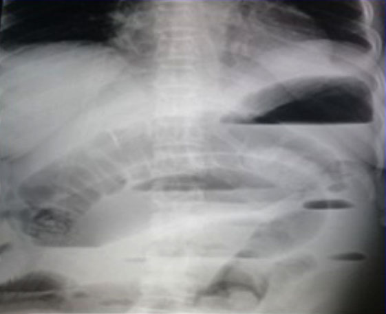
Plain radiographs of the abdomen showed distended small bowel with air fluid levels as it is shown in Figure 1. Ultrasound scan of the abdomen revealed moderate amount of free fluid and empty uterus. Her laboratory workup revealed elevated white blood count and elevated white blood cell count of 16.9×10³/μL, hemoglobin was 8.8g/dL; severe electrolyte imbalance with serum potassium of 6.4 mmol/L (normal 3.5–5) and blood glucose level of 5.2 mol/dL. She had evidence of severe metabolic acidosis with serum pH of 7.18 (normal 7.36–7.44), hypoxia with pO2 of 80 mmHg (normal 85–105). The surgical and obstetric teams in the emergency room evaluated the patient. Two hour on resuscitation the patient’s urine output was 30 mL/hour, the BP 117/73 mmHg, PR 103 beats/min, and RR 22 bpm; the patient gained conscious.
The patient was taken up for emergency laparotomy. Intra-operatively findings were 1200 mL of hemorrhagic fluid which was suctioned from the abdomen, the ileum was wrapping clockwise around the sigmoid causing double closed loops IO leading to ischemia of both ileum and sigmoid colon. The ileum involved (40 cm length) was friable, and gangrenous but the sigmoid colon was edematous but viable. No perforations were identified; the ileum was mobilized and resected, end-to-end anastomosis of the ileum was done and the colon was detorted and left free. The abdominal ravage and insertion of abdominal drainage were done; the abdomen was closed in layers. The operation duration was 2 hours, at the end of operation the patient was relatively stable. She was admitted in ICU.
Post-operatively, she was kept on broad-spectrum antibiotics and IV fluids. Within the first 24 hours, the patient was relatively improving with relative stable vital signs. However, she developed pulmonary function deteriorated with development of respiratory distress syndrome, dropping of blood pressure, rising of pulse rate, and fever (39°C) on which the titration of vasoconstrictor was added to increase vascular tone and cardiac output, also ventilator support was used. Despite the effort to rescue her life, she continued to deteriorating to worse condition and eventually she expired on the third day post-operative due to overwhelming sepsis and organ dysfunction.
DISCUSSION
Ileosigmoid knotting is rare in pregnancy with only 14 reported cases up to 2017 [5]. The reason of rarity may be caused by under reported cases [6]. It is a compound IO where the small bowel commonly the ileum around the base of sigmoid causing double closed loop bowel obstruction and causing vascular strangulation [5],[6]. Ileosigmoid is categorized into three types: Type I is the most common (53.9–57.5%) type of ISK. In this type, ileum is an active component wrapping itself around the sigmoid colon (passive component) in the clockwise (type) or anticlockwise direction (type IB). Type II is the second common (18.9–20.6%) type of ISK, where the sigmoid colon is active component where it wraps itself around a loop of the ileum (passive component) in the clockwise or anticlockwise direction. Lastly, type III is less common (1.5%) type of ISK, where the ileo-cecal segment (active component) wraps itself around the sigmoid colon passive component [1],[2],[7],[8]. In our case the patient had Type1A ISK.
In pregnancy, most of the ISKs reported are multiparous in third trimester (4–6). Our patient was, gravida 10, para 8 at 38 weeks, and hence she was in risked population. However, other scholars have reported that sigmoid volvulus and ISK in early pregnancy [8],[9],[10].
Concurrently, ISK is highly associated with multiparity due to the fact that the condition is associated with laxity and decreased tone of anterior abdominal wall [6] in addition to the increased intra-abdominal pressure caused by increased gravid uterus in third trimester, hence the likelihood of volvulus and ISK to occur [7],[10].
Ileosigmoid knotting mortality rate varies from 0% to 48% [2]. The mortality rates are often related to the duration of the symptoms, presence or absence of gangrene, and general status of the patient involving the presence of septic shock [4],[9]. Timely diagnosis and early surgical intervention are predictors of favorable outcomes [7],[9]. In our patient, there was a delay on both diagnosis and definitive management as she arrived to the primary hospital (district hospital) two days after onset of clinical symptoms. But additional delay was attributed by long driving distance (121 km) from the district hospital; the condition became fatal. The knot was so repetitively twisted but not tightly to the sigmoid loop and that’s why it was not gangrene-like ileum loop.
Resuscitation by IV fluid to restore fluid loss, correction of electrolyte imbalance, prophylactic antibiotics, and nasogastric for abdominal decompression is the principal initial treatment in all patients with IO [5],[6]. Considering our patient who was already with toxic and septic gangrenous small bowel, the definitive surgical intervention was inevitable and simple sigmoid detorsion was preferred on addition to ileum resection and primary end to end anastomosis. Other procedures recommended in the literatures are sigmoidostomy or sigmoidopexy if the colon is viable, however the simple detorsion is preferred [6].
The prognosis for patients with ISK during pregnancy is poor [11], as it occurred in our case. While the major causes of maternal mortality are toxic, hypovolemic, and septic shock [1],[3]. Intrauterine fetal death is likely to be caused by impairment of placental blood flow due to increased intra-abdominal pressure [6].
CONCLUSION
Ileosigmoid knotting in pregnancy is a rare disease which normally occurs in multiparous women in the third trimester. The presence of a clinical cardinal feature for IO; severe abdominal pain, distension, and absolute constipation in a pregnant patient may be suggestive of IO. On the other hand, ISK may be difficult at primarily health center level due to inexperience staff. However, good health policy and referring system may help to rescuer the patient’s lives. Since, the clinical appearance of the disease in pregnant patients is not distinctive from that of no pregnant women, the accurate preoperative diagnosis is not easy because the disease is rare and diagnostic radiographic studies are last option in pregnant women. Furthermore, the patients are usually diagnosed with a non-specific acute abdomen. After effective resuscitation, emergency surgery should be performed. Ileum resection with primary anastomosis and sigmoid resection with diverting colostomy are preferred in cases of bowel gangrene, whereas simple detorsion is used in non-gangrenous cases. However, regardless of all treatment strategies and approach, prognosis of ISK in pregnancy has poor prognosis to both maternal and fetus.
REFERENCES
1.
Li X, Zakariah SM, Shi Y. A case of ileosigmoid knotting in a Ghanaian patient. Int J Gen Med 2020;13:1265–9. [CrossRef]
[Pubmed]

2.
Shuaib A, Khairy A, Aljasmi M, Sallam MA, Abdulsalam F. Ileosigmoid knotting: A rare cause of intestinal obstruction and bowel ischemia—Case report with literature review. Open Access Emerg Med 2020;12:155–8. [CrossRef]
[Pubmed]
3.
Abebe K, Sherefa K, Teshome H, Abebe E. Ileosigmoid knotting: Analysis of patients clinical profiles and determinants of outcomes. Surg Res Pract 2020;2020:3826138. [CrossRef]
[Pubmed]
4.
Mutiibwa D, Tumusiime G. Ileosigmoid knotting in pregnancy: A case report seen in Uganda. East Cent Afr J Surg 2013;18(3):98–100.
5.
Mlambo BR, Mungazi SG, Maunganidze AJV. Ileosigmoid knotting in pregnancy: A case report from Zimbabwe. East Cent Afr J Surg 2017;22(2):76–80. [CrossRef]
6.
Atamanalp SS. Ileosigmoid knotting in pregnancy. Turkish J Med Sci 2012;42(4):553–8. [CrossRef]
7.
ToktaşO, Çim N. Ileosigmoid knot as a cause of acute abdomen at 28 weeks of pregnancy: A rare case report. East J Med 2017;22(3):110–3. [CrossRef]
8.
Anajjar M, El Brahmi Y, El Mouhafid F, et al. Ileosigmoid knot during pregnancy: Unusual cause of intestinal obstruction. PAMJ Clinical Medicine 2020;3(54):1–5.
9.
Karuhanga TA, Lulengo JM. Sigmoid volvulus in second trimester is challenge to obstetric and surgical departments at St. Francis Referral Hospital, Kilombero, Tanzania. Clin Med Rev Case Rep 2020;7(9):319. [CrossRef]
10.
Shimizu R, Hoshino Y, Irie H, et al. Ileosigmoid knot at week 13 of pregnancy: Report of a case. Int Surg 2014;99(3):230–4. [CrossRef]
[Pubmed]
11.
Maunganidze AJV, Mungazi SG, Siamuchembu M, Mlotshwa M. Ileosigmoid knotting in early pregnancy: A case report. Int J Surg Case Rep 2016;23:20–2. [CrossRef]
[Pubmed]
SUPPORTING INFORMATION
Acknowledgments
The authors would like to acknowledge the great support from the doctors and nurses from St. Francis Referral and Mulimba District Hospitals, respectively, and other staff members for patient care and extra patient’s information.
Author ContributionsTheresia Andrew Karuhanga - Substantial contributions to conception and design, Drafting the article, Final approval of the version to be published
Fassil Gebreegziabher Tekie - Substantial contributions to conception and design, Drafting the article, Final approval of the version to be published
Balichene Philbert Madoshi - Substantial contributions to conception and design, Drafting the article, Final approval of the version to be published
Guaranter of SubmissionThe corresponding author is the guarantor of submission.
Source of SupportNone
Consent StatementWritten informed consent was obtained from the patient for publication of this article.
Data AvailabilityAll relevant data are within the paper and its Supporting Information files.
Conflict of InterestAuthors declare no conflict of interest.
Copyright© 2022 Theresia Andrew Karuhanga et al. This article is distributed under the terms of Creative Commons Attribution License which permits unrestricted use, distribution and reproduction in any medium provided the original author(s) and original publisher are properly credited. Please see the copyright policy on the journal website for more information.


