 |
|
Clinical Image
| ||||||
| Klippel–Trenaunay syndrome | ||||||
| Kamal Kant1, Richa Purohit2 | ||||||
|
1MS, FACS, FRCS (Glasgow, UK), Sr. Consultant, Surgery, Medipulse Hospital, Jodhpur, Rajasthan, India 342003, B 24, Shastri Nagar, Jodhpur, Rajasthan, India 2MS, MRCS (UK), Consultant Surgery, Dr. S.N. Medical College, Jodhpur, Rajasthan, India | ||||||
| ||||||
|
[HTML Abstract]
[PDF Full Text]
[Print This Article] [Similar article in PubMed] [Similar article in Google Scholar] |


| How to cite this article |
| Kant K, Purohit R. Klippel–Trenaunay syndrome. Edorium J Surg 2018;5:100027S05KK2018. |
|
CASE REPORT
| ||||||
|
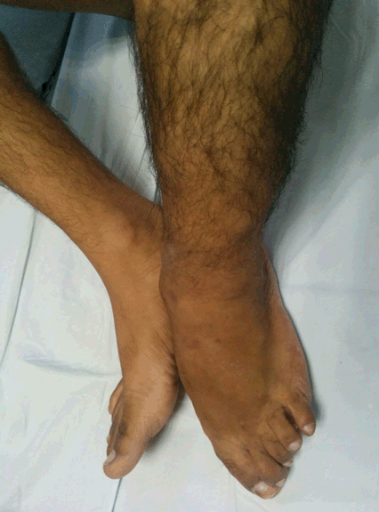
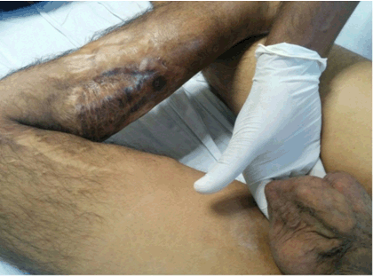
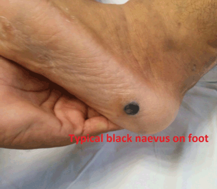
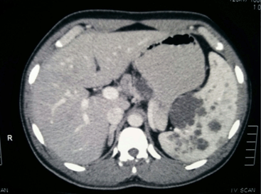
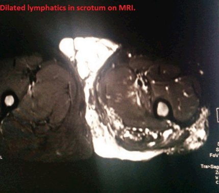
A 17-year-old male with history of repeated surgeries for cystic lesions in neck and lower limb since infancy, abnormal overgrowth of toes and limbs, anaemia requiring multiple transfusions. On examination, young male, anaemic short statured with lower limb disabilities (Figure 1). Abdominal examination revealed huge splenomegaly, no ascitis, no other organomegaly. Scrotum contained soft cystic swelling bilaterally (Figure 2) transilluminant and no impulse. Left lower leg was deformed with scars all over as result of previous surgeries, overgrowth of toes and black naevus on foot (Figure 3). Patient was evaluated biochemically and by imaging USG, MRI of abdomen. MRI images (Figures 4, Figure 5 , Figure 6, Figure 7) confirmed the diagnosis of Klippel–Trenaunay syndrome. Patient underwent splenectomy and was in follow up for one year. Keywords : Cystic swelling, Genetic mutation, Klippel–Trenaunay syndrome | ||||||
| ||||||
| ||||||
| ||||||
| ||||||
| ||||||
| ||||||
| ||||||
DISCUSSION | ||||||
|
The syndrome is a genetically inherited condition affecting the development of blood vessels, soft tissues and bones. It affects 1 in 100,000 populations [1] all over the world. It is caused by genetic mutation most commonly affecting PIKCA gene which is responsible for development of tissues in the body resulting in overgrowth. Common presenting features included:
| ||||||
|
CONCLUSION
| ||||||
|
This syndrome is a genetically inherited rare condition affecting malformation of soft tissue including blood vessels, bones, lymphatics in limbs and viscera. No definite treatment is available.
| ||||||
|
REFERENCES
| ||||||
| ||||||

|
[HTML Abstract]
[PDF Full Text]
|
|
Author Contributions
Kamal Kant – Substantial contributions to conception and design, Acquisition of data, Analysis and interpretation of data, Drafting the article, Revising it critically for important intellectual content, Final approval of the version to be published Richa Purohit – Substantial contributions to conception and design, Acquisition of data, Analysis and interpretation of data, Drafting the article, Revising it critically for important intellectual content, Final approval of the version to be published |
|
Guarantor of Submission
The corresponding author is the guarantor of submission. |
|
Source of Support
None |
|
Consent Statement
Written informed consent was obtained from the patient for publication of this clinical image. |
|
Conflict of Interest
Author declares no conflict of interest. |
|
Copyright
© 2018 Kamal Kant et al. This article is distributed under the terms of Creative Commons Attribution License which permits unrestricted use, distribution and reproduction in any medium provided the original author(s) and original publisher are properly credited. Please see the copyright policy on the journal website for more information. |
|
|