|
Case Report
Bouveret’s syndrome as a rare cause of gallstone ileus: A surgical challenge
1 MD, General surgeon, Bariatric surgeon, Division of General Surgery/Laparoscopy, Centro Médico de Especialidades, Ciudad Juárez, Chihuahua, Mexico
2 MD, General surgeon, Division of General Surgery/Laparoscopy, Centro Médico de Especialidades, Ciudad Juárez, Chihuahua, Mexico
3 MD, General physician, Instituto de Ciencias Biomédicas, Universidad Autónoma de Ciudad Juárez, Ciudad Juárez, Chihuahua, Mexico
4 MD, General physician, Centro Quirúrgico de Lourdes, Ciudad Juárez, Chihuahua, Mexico
5 MD, General physician, Universidad Autónoma de Guadalajara, Zapopan, Jalisco, Mexico
Address correspondence to:
Danai Hernández Carreón
Vicente Guerrero 8907, Fraccionamiento Las Quintas, CP 32401, Ciudad Juárez, Chihuahua,
Mexico
Message to Corresponding Author
Article ID: 100065S05CA2026
Access full text article on other devices

Access PDF of article on other devices


How to cite this article
Avilez CTP, Cereceres SJR, Jaime CAL, Carreón DH, García AGA, De La Garza ASP. Bouveret’s syndrome as a rare cause of gallstone ileus: A surgical challenge. Edorium J Surg 2026;12(1):1–5.ABSTRACT
Introduction: Bouveret syndrome, a rare cause of gastrointestinal outlet obstruction, was first described by Beassier in 1770. Afterward, this condition was named after Leon Bouveret in 1896. Bouveret syndrome is characterized by the passage of a gallstone through a cholecystogastric or more commonly through a cholecystoduodenal fistula, causing obstruction.
Case Report: A 70-year-old male patient without chronic degenerative diseases and with history of epigastric pain months prior to his current evaluation. Five days before hospital admission, the patient experienced worsening of the epigastric pain with an intensity of 8/10 accompanied by anorexia, vomiting, jaundice, and choluria. Physical examination with evidence of generalized jaundice ++, painful and distended abdomen in the upper right quadrant. Diagnostic protocol was initiated based on preoperative tests, which showed hyperbilirubinemia of 2.78 mg/dL with an obstructive pattern and alteration of liver function tests. The hepatobiliary ultrasound showed a lithiasic sclero-atrophic gallbladder, a common bile duct diameter of 5 mm and positive sonographic murphy sign. The patient was taken to the operating room where, after general anesthesia and in a supine position, a 13 mmHg pneumoperitoneum was created and laparoscopic working ports were introduced. A laparoscopic cholecystectomy + trancystic cholangiography + resection of the cholecystoduodenal fistula were performed.
Conclusion: A high degree of clinical suspicion and imaging studies are required to establish Bouveret syndrome diagnosis, nevertheless in 50% of cases, the diagnosis is made intraoperatively. Early diagnosis and adequate surgical intervention are essential for a favorable prognosis.
Keywords: Cholecystoduodenal fistula, Cholelithiasis, Gallstone ileus, Pneumobilia
INTRODUCTION
Bouveret syndrome, a rare cause of gastrointestinal outlet obstruction, was first described by Beassier in 1770. Afterward, this condition was named after Leon Bouveret in 1896, who presented two cases of patients with this condition. Bouveret syndrome is caused by the impaction of a gallstone into the pylorus or duodenum and is characterized by the passage of a gallstone through a cholecystogastric or more commonly, through a cholecystoduodenal fistula, causing obstruction of the gastric outlet. Cholelithiasis is a common disease which can lead to several complications. Bouveret syndrome accounts for 0.3% of complications from cholelithiasis, and gallstone ileus accounts for approximately 1–3% of cases of mechanical gastrointestinal obstruction. The typically affected population is the elderly, with a median age of 70 years old, which contributes to the high mortality rates estimated to be 12–30% [1],[2],[3],[4].
CASE REPORT
A 70-year-old male patient without chronic degenerative diseases and with history of epigastric pain months prior to his current evaluation. Five days before hospital admission, the patient experienced worsening of the epigastric pain with an intensity of 8/10 accompanied by anorexia, nausea, vomiting, as well as jaundice and choluria. Physical examination with evidence of generalized jaundice ++, painful and distended abdomen in the upper right quadrant with no evidence of peritoneal irritation.
Diagnostic protocol was initiated based on preoperative tests, which showed leukocytes of 7.33, platelets of 254,000, total bilirubin of 2.78 mg/dL, direct bilirubin of 1.56 mg/dL, indirect bilirubin of 1.22 mg/dL, aspartate aminotransferase of 126 U/L, alanine aminotransferase of 333 U/L, gamma glutamyl transpeptidase of 749 U/L, alkaline phosphatase of 144 U/L, and lactate dehydrogenase of 233 U/L. The hepatobiliary ultrasound showed a lithiasic sclero-atrophic gallbladder, a common bile duct diameter of 5 mm and positive sonographic murphy sign.
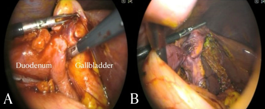
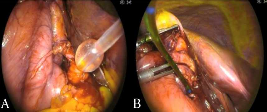
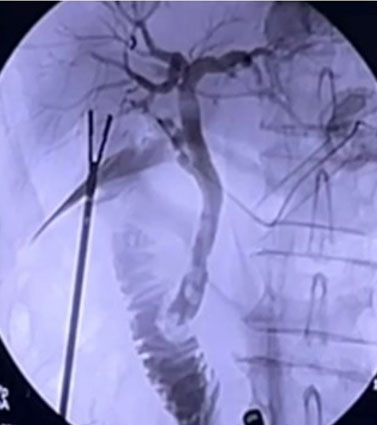
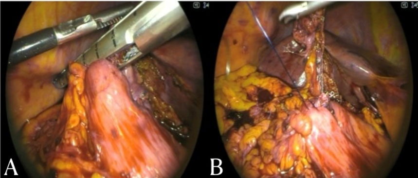
The patient was taken to the operating room where, after general anesthesia and in a supine position, a 13 mmHg pneumoperitoneum was created and laparoscopic working ports were introduced. During diagnostic laparoscopy a cholecystoduodenal inflammatory process was identified, as well as, a cholecystoduodenal fistula (Figure 1). The fistula was dissected, followed by the dissection of the Calot’s triangle, dissecting the cystic duct and performing transcystic bile duct exploration, identifying a stone within. A balloon catheter was introduced and gallstones were extracted (Figure 2). A transcystic cholangiography was performed, showing adequate passage into the duodenum and into the right, left, and common hepatic ducts (Figure 3). The balloon catheter was removed, the cystic duct and artery were stapled and the gallbladder was dissected. Then, the cholecystoduodenal fistula was stapled with a tri-staple stapler and reinforced with mechanical sutures (Figure 4). Finally, hemostasis was confirmed, a thorough cleaning was performed, a Penrose drain was placed in Winslow’s hiatus and the gallbladder was extracted. A laparoscopic cholecystectomy + transcystic cholangiography + resection of the cholecystoduodenal fistula were performed. The patient presented favorable clinical evolution and was discharged three days after surgery.
DISCUSSION
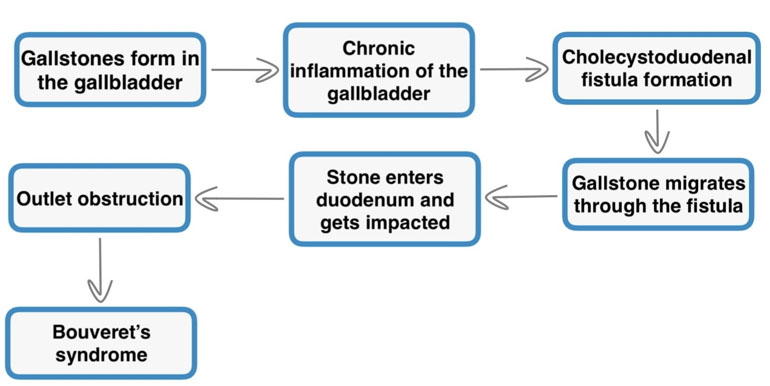
The inflammation and adherence of the gallbladder to the pylorus or duodenum are a consequence of multiple episodes of acute cholecystitis which can lead to an ischemic tear of the gallbladder and enteric wall, in addition to the mechanical tension exerted by the gallstone, all favoring the formation of a bilioenteric fistula (Figure 5). Alternatively, there have been reports of the development of bilioenteric fistulas secondary to gallbladder malignancy. Bilioenteric fistulas most commonly present as cholecystoduodenal fistulas, representing 68% of all bilioenteric fistulas, followed by cholecystocolic fistulas with 17% and lastly, by cholecystogastric fistulas, representing 5% of all cases, which is the rarest due to gastric wall thickness [1],[4],[5].
Bouveret’s syndrome risk factors include being elderly with a median age of 70 years, female sex, gallstones between 2 and 8 cm, and gastrointestinal anatomical alterations. The clinical presentation of Bouveret syndrome is nonspecific, which delays the diagnosis and contributes to high morbidity and mortality, in addition to associated comorbidities. In most cases in which a bilioenteric fistula is present, gallstones are eliminated through vomiting or defecation, and therefore, symptoms arise with larger stones. The most common symptoms are nausea, vomiting, hematemesis, weight loss, abdominal pain and anorexia. Hematemesis results from a Mallory–Weis tear and in some cases emesis can be severe enough that may lead to a Boerhaave syndrome. Physical examination may reveal abdominal distension and tenderness, as well as altered bowel movements. Bouveret’s syndrome complications include biliary or duodenal perforations, biliary infection, acute pancreatitis, cancer, and recurrent Bouveret syndrome [1],[4],[5],[6],[7].
Regarding the diagnosis, laboratory tests are non-specific. Bouveret syndrome is mainly diagnosed by imaging studies. Rigler’s triad, which consists of pneumobilia, an ectopic gallstone, and distended stomach, is pathognomonic, although it only appears in 35% of the cases. Abdominal ultrasound can also demonstrate Rigler’s triad. Esophagogastroduodenoscopy provides direct visualization of the gallstones and has the additional advantage of being therapeutic, however, in almost 33% of the cases, it fails to visualize the gallstone. The best diagnostic method is the contrast-enhanced computed tomography, which has a sensitivity of 90% and specificity of 100% in detecting gallstone ileus. Abdominal computed tomography (CT) can provide more detailed information about the fistulous tract, and the ectopic calculus, and may help identify complications [4],[7],[8].
Treatment options include endoscopic, laparoscopic, or open disimpaction of the stone. The endoscopic approach, diagnostic and potentially therapeutic, is preferred due to the lowest mortality; nonetheless, its effectiveness depends on the size of the gallstone and on adequate endoscopic skills. Endoscopic modalities include nets, baskets, and lithotripsy techniques such as mechanical, electrohydraulic, and intracorporeal lasers which can be used prior to endoscopic extraction. Complications of endoscopic treatment include impaction of the stone in the esophagus, gastrointestinal bleeding, and perforation [2],[6],[9].
Although the endoscopic approach is preferred, surgery will be required in 90% of cases. There are currently 3 surgical approaches. Laparoscopic or open enterotomy with gallstone extraction and spontaneous closure of the fistula, which should be offered in elderly patients with associated comorbidities. A one stage procedure involving enterotomy and gallstone extraction, with or without cholecystectomy and repair of the fistula, which should be considered in patients with good general condition. The last one is the two stage procedure consisting of enterotomy with gallstone extraction followed by an elective cholecystectomy and fistula repair after 4–6 weeks. Cholecystectomy is indicated to prevent recurrence and biliary complications, and only 10% of the patients will require delayed cholecystectomy with fistula repair for persistent symptoms. Laparoscopic surgery is a safe and an effective alternative to open surgery for gallstone ileus; however, it is used in only 10% of surgical managed gallstone ileus cases with a high conversion rate to laparotomy [2],[3],[6].
CONCLUSION
Due to the non-specific symptoms and low incidence of the syndrome, a high degree of clinical suspicion and imaging studies are required to establish Bouveret syndrome diagnosis, nevertheless, in 50% of cases the diagnosis is made intraoperatively. Early diagnosis and adequate surgical intervention are essential for a favorable prognosis. The endoscopic approach remains the first-line treatment, particularly in the elderly with comorbidities. Alternatively, the laparoscopic approach and repair of the fistula remains a complex and controversial issue among surgeons.
REFERENCES
1.
Thatipalli N, Gattani R, Nayak K, Sudabattula K. Bouveret syndrome: Etiology, clinical presentation, differential diagnosis, complications, and treatment options. Cureus 2024;16(7):e64754. [CrossRef]
[Pubmed]

2.
Evola G, Caramma S, Caruso G, Dapri G, Evola FR, Reina C, et al. Bouveret’s syndrome as a rare complication of cholelithiasis: Disputes in current management and report of two cases. Int J Surg Case Rep 2020;71:315–8. [CrossRef]
[Pubmed]
3.
Atri S, Elaifia R, Sebai A, Hammami M, Haddad A, Kacem JM. Bouveret syndrome: A rare form of gallstone ileus a case report. Int J Surg Case Rep 2024;116:109438. [CrossRef]
[Pubmed]
4.
Osman KT, Wahba A, Farhoud A, Abdekfattah AM, Maselli DB. Bouveret’s syndrome: An extensive summary of the literature. Archives of Gastroenterology Research 2020;1(2):40–46.
5.
Ignjatovic NS, Golubovic ID, Djordjevic MN, Stojanovic MM, Benedeto Stojanov DA, Ignjatovic JS, et al. Bouveret’s syndrome as a rare life-threatening complication of gallstone disease—A surgical problem: Two case reports. Medicina (Kaunas) 2024;61(1):5. [CrossRef]
[Pubmed]
6.
Singh G, Merali N, Shirol S, Drymousis P, Singh S, Veeramootoo D. A case report and review of the literature of Bouveret syndrome. Ann R Coll Surg Engl 2020;102(1):e15–9. [CrossRef]
[Pubmed]
7.
Sabir A, Mushtaq R, Arshad R, Khalid N, Ayub M, Maqbool S, et al. Bouveret’s syndrome: A rare case of gallstone causing gastric outlet obstruction. Clin Case Rep 2024;12(6):e8969. [CrossRef]
[Pubmed]
8.
Navarro-Del Río E, Hernández-Zúñiga JF. Bouveret’s syndrome: A rarest complication of cholelithiasis. A case report and literature review. Cir Cir 2020;88(1):95–99. [CrossRef]
[Pubmed]
9.
Khan A, Khan T, Mushtaq K, Zelt C, Sharma N. Bouveret syndrome: A rare cause of gastric outlet obstruction and treatment options. Cureus 2025;17(1):e77032. [CrossRef]
[Pubmed]
SUPPORTING INFORMATION
Acknowledgments
We thank the staff of Centro Médico de Especialidades for their constant drive to learn.
Author ContributionsCarlos Tadeo Perzabal Avilez - Substantial contributions to conception and design, Analysis of data, Interpretation of data, Revising it critically for important intellectual content, Final approval of the version to be published
Saul Jasam Ruiz Cereceres - Substantial contributions to conception and design, Analysis of data, Revising it critically for important intellectual content, Final approval of the version to be published
Cesar Alberto Lopez Jaime - Substantial contributions to conception and design, Analysis of data, Interpretation of data, Revising it critically for important intellectual content, Final approval of the version to be published
Danai Hernández Carreón - Substantial contributions to conception and design, Acquisition of data, Analysis of data, Interpretation of data, Drafting the article, Revising it critically for important intellectual content, Final approval of the version to be published
Aldo Gustavo Acosta García - Substantial contributions to conception and design, Analysis of data, Revising it critically for important intellectual content, Final approval of the version to be published
Ana Sofía Perzabal De La Garza - Substantial contributions to conception and design, Acquisition of data, Analysis of data, Revising it critically for important intellectual content, Final approval of the version to be published
Guaranter of SubmissionThe corresponding author is the guarantor of submission.
Source of SupportNone
Consent StatementWritten informed consent was obtained from the patient for publication of this article.
Data AvailabilityAll relevant data are within the paper and its Supporting Information files.
Conflict of InterestAuthors declare no conflict of interest.
Copyright© 2026 Carlos Tadeo Perzabal Avilez et al. This article is distributed under the terms of Creative Commons Attribution License which permits unrestricted use, distribution and reproduction in any medium provided the original author(s) and original publisher are properly credited. Please see the copyright policy on the journal website for more information.